Most genetic diseases are caused by a defective gene, which results in a loss of activity of some enzymes. Even though the body has thousands of enzymes, the loss of only one may be disastrous. The most common genetic disease in the United States is a cystic fibrosis, which affects 1 in 2000 births, although approximately 1 in 20 persons may carry the genetic trait. Although lung function appears normal at birth, early in a childhood a thick mucus develops and results bronchiolar obstruction. Infection occurs and becomes difficult to eradicate, even with antibiotics.
When such an infection develops in the lungs, the subsequent inflammatory reaction results in the destruction of bacteria, leukocytes, and tissue. The process of cellular destruction releases DNA, which in turn substantially increases the viscosity of the mucus. An experimental treatment for cystic fibrosis involves the use of DNase, an enzyme that degrades extracellular DNA but has no effect on the DNA within intact cells. DNAse is produces by recombinant DNA technology and is composed of a single chain of 260 amino acids.
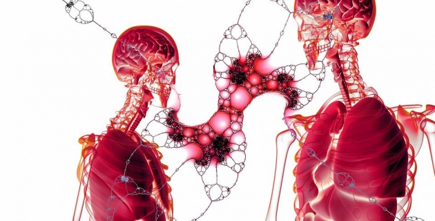
Image credits: Geralt via Pixabay
Another genetic disease, phenylketonuria, affects 1 in 10,000; most states now require screening of newborn infants for this disease. Tay-Sachs disease affects 1 in 1000 among persons of eastern European Jewish origin. One out of every 10 black people in the United States carries the gene for sickle cell disease.
In almost every ethnic, demographic, or racial group certain genetic diseases occur at much higher frequencies among their members than in the general population. Paget's disease, a disease of the bones, occurs frequently among people of the English decent. Polynesians are prone to have clubfoot, and Finns tend to have a kidney disease. There is a higher than average frequency of deafness in Eskimos and of glaucoma in Icelanders. Native Americans and Mexicans are very susceptible to diabetes and gallbladder disease. Impacted wisdom teeth occur frequently among Europeans and Asians but rarely among Africans. However, impacted wisdom teeth may also be dietary in origin. Today there are at least 1200 distinct inherited genetic diseases that have been identified.
Sickle Cell Anemia
Each of these amino acids was designated by a certain arrangement of three nucleotide in mRNA. Altogether, there must have been 438 nucleotide, arranged in the proper sequence in order to form this molecule of hemoglobin. Look at the amino acid 6, the one in color. This amino acid, Glue, is a glutamic acid. The mRNA code group for glutamic acid is either GAA or GAG. If the middle codon of this group is changed from A to U, the sequence becomes either GUA and GUG, both of which designate the amino acid valine (Val). That is, if there is a change in only one of the chain of hemoglobin, a different type of molecule is produced. This type of hemoglobin is called S and causes the genetic disease sickle cell anemia.
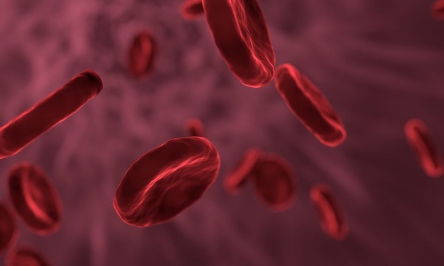
Image credits: Allinonemovie via Pixabay
The red blood cells normally have a concave shape when deoxygenated. It contains hemoglobin S look like a sickle. These sickle cells are more fragile than normal red blood cells, leading to anemia. They can also occlude capillaries, leading to thrombosis. The points and abnormal shapes of the sickle cells cause slowing and sludging of the red blood cells in the capillaries, with resulting hypoxia of the tissues. This produces such symptoms as fever, swelling, and pain in various parts of the body. Eventually the spleen is affected. Many victims of severe sickle cell anemia die in childhood.
Sickle cell anemia is a hereditary condition found primarily among Hispanics and blacks. Many of these people have the sickle cell trait but are relatively unaffected by it until there is a sharp drop in blood oxygen level, such as might be caused by strenuous exercise at high altitudes, underwater swimming, and alcohol intoxication.
Phenylketonuria
Phenyketonuria (PKU) results when the enzyme phenylalanine hydroxylase is absent. A person with PKU cannot convert phenylalanine to tyrosine, and so the phenylalanine accumulates in the body, resulting in injury to the nervous system. In infants and in children up to age 6, an accumulation of phenylalanine leads to retarded mental development.
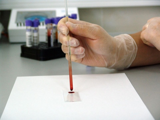
Image credits: PublicDomainPictures via Pixabay
The disease can be readily diagnosed from a sample of blood or urine. Treatment consists of giving affected person a diet low in phenylalanine and adding tyrosine to the diet. Tyrosine is essential in the absence of phenylalanine.
Signs and Symptoms
Untreated PKU can lead to intellectual disability, seizures, behavioral problems, and mental disorders. It may also result in a musty smell and lighter skin. Babies born to mothers who have poorly treated PKU may have heart problems, a small head, and low birth weight.
Because the mother's body is able to break down phenylalanine during pregnancy, infants with PKU are normal at birth. The disease is not detectable by physical examination at that time, because no damage has yet been done. Newborn screening is performed to detect the disease and initiate treatment before any damage is done. The blood sample is usually taken by a heel prick, typically performed 2-7 days after birth. This test can reveal elevated phenylalanine levels after one or two days of normal infant feeding.
Read more on this source.
Galactosemia
Galactosemia results from the lack of the enzyme uridyl transferase, which catalyzes the formation of the glucose from galactose. This disease may result in an increased concentration of galactose in the blood. Galactose in the blood is reduced in the eye to galacticol, which accumulates and causes a cataract. Ultimately, if galactose continues to accumulate, liver failure and mental retardation will occur. This disease can be controlled by the administration of a diet free of galactose.
Wilson's Disease
In this disease, copper accumulates in the liver, kidneys, and brain. There is also an excess of copper in the urine. If deposition of copper in the liver becomes excessive, cirrhosis may develop. In addition, accumulation of copper in the kidneys may lead to damage of the renal tubules, leading to increased output of amino acids and peptides.
Albinism

Image credits: Babbur via Pixabay
Albinism is caused by the lack of the enzyme tyrosinase, which is necessary for the formation of melanin, the pigment of the hair, skin, and eyes. Consequently, albinos have very white skin and hair. Although this disease is not serious, persons affected by it are very sensitive to sunburn.
Hemophilia

Image credits: Whitesessions via Pixabay
Hemophilia is caused by a missing protein, an antihemophilic globulin, which is important in the normal clotting process of the blood. Consequently, any cut may be life threatening to hemophiliacs, but the primary damage is the crippling effect of repeated episodes of internal bleeding into body joints.
Muscular Dystrophy
Courtesy of the video: NationwideChildrens via Youtube
One form of muscular dystrophy, Duchenne's muscular dystrophy, is caused by a lack of protein caller dystrophin. This disease primarily affects the boys and causes progressive weakness and muscle wasting of muscles. Victims are usually confined to a wheelchair by age 12 and die before age 30 because of respiratory failure. Currently, there is no treatment for this disease by the new discovery of stem cell therapy is hopefully a cure.
Dystrophin is a large protein with molecular mass of 400,000. It is normally found in the muscle cell membrane, on the side facing the cell interior.
Researchers are investigating many details about satellite cells and the causes of muscle damage as well as treatments that help reduce muscle damage, such as anti-inflammatory treatments.
Studies are examining ways to preserve, and possibly restore, muscle function by transplanting dystrophin-producing cells into patients. These cells could be healthy donors cells or a patient's own cells that have been genetically modified.
Induced pluripotent steem cells (iPSCs) are being studied as an option for making large numbers of cells with healthy dystrophin genes.
Read more on this source.
Niemann-Pick disease
This disease is caused by a lack of the enzyme sphingomyelinase, which causes an accumulation of sphingomyelin in the liver, spleen, bone marrow, and lymph nodes. This disease affects the brain and causes mental retardation and early death.
Gaucher's disease
It is caused by a lack of the enzyme glucocerebrosidase, which is necessary for the cleavage of glucocerebrosides into glucose and ceramide. This disease is characterized by the accumulation of glycolipids in the spleen and liver. In children, Gaucher's disease causes severe mental retardation and early death. In adults, the spleen and liver enlarge progressively but the disease is compatible with long life.
Tay-Sachs
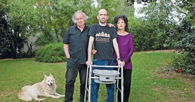
Oded Stern, a patient of Tay-Sachs discovered his disease at the age of 29 and suffers brain defects. His dad is courageous to seek the treatment. (Image credits)
It is due to a lack of the enzyme hexosaminidase. A leading to the accumulation of glycolipids in the brain and in the eyes. Red spots show up in the retina, and there is also muscular weakness. This disease is fatal to infants before the age of four.
Krabbe's
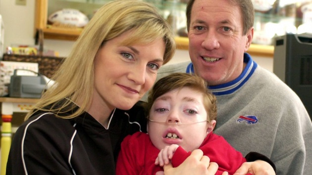
Due to lack of enzyme galactosidase, it is characterized by mental retardation and the absence of myelin. In the picture above is Hunter who passed away in 2005. He died at the age of 8. (Image credits: NFR)
Farber's
It is due to lack of enzyme ceramidasse, is characterized by hoarseness, dermatitis, skeletal degeneration, and mental retardation.
Lou Gehrig's Disease

In the picture is Stephen Hawking who survived 55 years of lived with ALS. (Image credits)
Amyotrophic lateral sclerosis, ALS, also known as Lou Gehrig's disease, is characterized by the degeneration of the nerves that control motor activity in the brain and spinal chord. This degeneration results in a progressive wasting of the muscles. Individuals with such a disease lose their ability to walk, talk, and swallow. Their intellect, however, is unimpaired. Most patients die within 2 to 5 years after the first symptoms, and death usually comes from suffocation.
Recent evidence has shown that patients with ALS have an accumulation of glutamate in the fluid between the cells in the brain. As a result of this accumulation, excess calcium ions flow into the nerve cells. The excess calcium ions, in turn, have a deadly effect on the neurons that control motor activity, and those cells soon die.
Physical activity
A 2005 systematic review found no relationship between the amount of physical activity and the risk of developing ALS, but did not find that increased leisure time physical activity was strongly associated with an earlier age of onset of ALS. A 2007 review concluded that physical activity was probably not a risk factor for ALS. A 2009 review found that the evidence for physical activity as a risk factor for ALS was limited, conflicting, and of insufficient quality to come to a firm conclusion. A 2014 review concluded that physical activity in general is not a risk factor for ALS, that soccer and American football are possibly associated with ALS, and that there was not enough evidence to say whether or not physically demanding occupations are associated with ALS. A 2016 review found the evidence inconclusive and noted that differences in study design make it difficult to compare studies, as they do not use the same measures of physical activity or the same diagnostic criteria for ALS.
Read more on this source.
Classification of Genetic Diseases
Genetic diseases may be classified into the three major categories: Chromosomal disorders, in which there is an excess of loss of chromosomes, deletion of part of a chromosome, or translocation or a chromosome. Examples include down's syndrome and chronic myelogenous leukemia, monogenic disroders, which involve one mutant gene, multifunctional disorders involve the action of a number of genes. One example is essential hypertension.
Treatment of Genetic Diseases
One of the following methods may be used in the treatment of genetic diseases.
1. Correct the metabolic consequences of the disease by supplying the missing product. For example, a patient with a familiar goiter can be treated with the missing hormone L-thyroxine. 2. Replace the missing enzyme or hormone. 3. Remove excess stored substance. Example: administration of Vitamin B12 in the treatment of methylmalonic acidemia. 3. Correct the major abnormality. One example is the use of a liver transplant in a patient with galactosema (primarily used on patients with advanced cases of this disease).
Recombinant DNA (Gene Splicing)
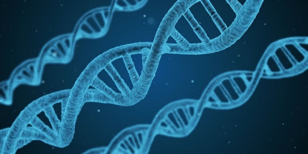
Image credits: Qimono via Pixabay
The term DNA refers to DNA molecules that have been artificially created by splicing segments of DNA from one organism into the DNA of completely different organism. Almost any organism (animal, plant, bacteria, virus) can serve as DNA donor. Theoretically any organism also can serve as DNA acceptor, but to date the bacterium Escherichia coli (E.coli) has been used almost exclusively as a DNA acceptor. E.coli has selected because its genetic makeup has been thoroughly studied. Most of E.coli's 300 to 4000 genes are contained within a large, single, ringed chromosomes. In addition to this large chromosome, there are plasmids, smaller closed loops of DNA consisting only a few genes. It is these plasmids that act as DNA acceptors for E.coli, since the plasmids are known to multiply independently within the host cell as the cell itself replicates.
How does gene splicing work? First, the E.coli is placed in a detergent solution to break upon the cells. The plasmids are treated with restriction enzymes, which cut the plasmid DNA at specific points. DNA from another organism (also cleaved by similar method) is inserted into E.coli. The new cells are isolated and are allowed to replicate (E.coli duplicates itself every 20 to 30 minutes). As the cells replicate, they synthesize the proteins coded for by their DNA, including those coded for by inserted DNA.
Recombinant DNA research has produced human insulin, interferon, growth hormone, somatostatin, and some vaccines. Recombinant DNA research has led two widely used drugs to treat lowered red blood cell levels in dialysis patients and neutropenia in chemotherapy patients. Recently, recombinant DNA products have been tried in patients with cystic fibrosis and emphysema. Theoretically, gene splicing could be used to find cures for genetic diseases and for cancer and to manufacture various enzymes, hormones, antibodies, and other substances that are difficult to isolate and produce pharmaceutically.
Opponents of recombinant DNA research point out considerable risks involved. They fear that the synthetic strain of E.coli might be accidentally released and cause a worldwide epidemic and the death of large populations. They also believe that this type of research might be used politically to control human behavior.
Genetic Markers
One of the newer topics of cancer research is genetic markers, defects in one part of a chromosome that is believed can cause a certain disease. A genetic marker of Alzheimer's disease has been found on chromosome 21, the same chromosome that is associated with Don's syndrome. Since a chromosome can include many, many genes, much work is yet to be done in this area.
Oncogenes
Oncogenes are genes that appear to trigger uncontrolled, or cancerous, growth. Cancerous cells exhibit three general characteristics:
- Uncontrolled growth
- Invasion of body tissues
- Spread to other body parts (metastasis)
In cancer, the genes controlling growth are abnormal, but little is known about how the cell growth is controlled. It is the second most common cause of death in the United States, after cardiovascular disease. Its incidence increase with age, and a wide variety of body organs can be affected. The abnormal production of enzymes, hormones, and proteins is frequently associated with cancer. These products are called tumor markers. measurement of such markers is helpful in diagnosing and treating cancer. For example, the detection of the enzyme prostatic acid phosphatase maybe associated with cancer of the prostate gland, and abnormal amounts of calcitonin may indicate carcinoma of the thyroid gland.

Image credits: Marijana1 via Pixabay
What causes cancer? It is believed that all cancer-causing agents can be grouped into three main categories: radiant energy, chemical compounds, and viruses. Radiant energy such as X-rays and Y-rays are carcinogenic because of the formation of *free radicals in the tissues. Radiation can also damage DNA and so is *mutagenic. Many chemical compounds in common use are known to be carcinogenic. As much as 75% of human cancers are caused by chemicals in the environment. Among these carcinogenic chemicals are benzene, asbestos, and fused-ring compounds such as benzopyrene found in cigarette smoke. Oncogenic viruses contain either DNA or RNA. Under certain circumstances, infection of appropriate cells with polyoma virus or SV 40 virus can result in a malignant transformation.
DNA Fingerprinting
Courtesy of the video: Bozeman Science via Youtube
Fingerprint analysis was and still is an effective method for identifying individuals since no two persons have the same fingerprints. However, modern genetic "fingerprinting" can accomplish the same thing since no two individuals have the same DNA unless they are identical twins. Samples of DNA can be taken from saliva, skin, hair, semen, or bone. With current laboratory procedures only small part of the DNA sequence is examined. The primary method used in DNA fingerprinting is called restriction fragment length polymorphism (RFLP). In this method, special enzymes are used to "cut" samples of DNA into sections called restrictions fragments, which are then separated by gel electrophoresis according to the size. The electrophoresis patterns are quite distinctive and when exposed to photographic. film produce a "fingerprint". A match can have an accuracy of 99%.
Samples too small to analyze by RFLP method can be identified by thee polymerase chain reaction (PCR) method, whereby the DNA is recopied many times until enough is present for a proper analysis. This method can be used to identify the DNA present in one tiny drop of blood or in one strand of hair.
-----------------------------------------------------------------------
All rights reserved 2019. No part of this article
may be reproduced without special credits
in writing from the publishers of Wikipedia, MayoClinic, and WebMD.


